A Silver Bullet for Cancer Treatment?
Nanoparticles may be able to tame an effective but damaging chemotherapy medication.
A Silver Bullet for Cancer Treatment?
Nanoparticles may be able to tame an effective but damaging chemotherapy medication.
Oncology nurses know the bright red drug “red devil” is a crucial weapon in the chemotherapy arsenal — a go-to for fighting breast, bladder and blood cancers.
Doxorubicin, the devil’s proper name, also is hazardous, blisters tissue on contact and causes side effects that range from mouth sores to congestive heart failure. The medication’s indiscriminate effects on healthy as well as cancerous tissue have earned it the grim nickname.
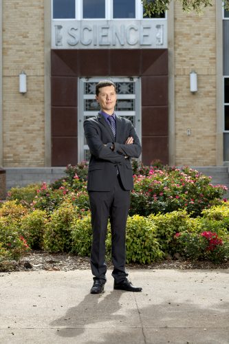
Anton Naumov studies how quantum dots can aid cancer-killing drugs without the extreme side effects. Photo by Jeffrey McWhorter
But a versatile material called graphene oxide might turn the doxorubicin sword into a precision scalpel — and help oncologists locate the tumor in the first place.
Anton Naumov, assistant professor of physics, is studying graphene oxide, a substance related to the graphite in pencils, generated in microscopic amounts. Naumov works with tiny pieces of this substance called quantum dots. Cheap, soluble and nontoxic, these nanoparticles might one day perform multiple cancer-related tasks by acting as a drug-delivery vehicle, imaging agent and noninvasive cancer sensor.
“We wanted to use the idea of integration to be able to utilize one platform, one nanomaterial for multiple roles,” Naumov said. He compared quantum dots’ potential to a cellphone’s ability to play videos and use GPS.
Graphene is a sheet of single-carbon atoms arranged in hexagons that resembles a tiled floor; a carpeting of oxygen molecules turns it into graphene oxide. A graphene quantum dot is a flake about 1/500th of a micrometer wide. By comparison, a human hair is roughly 80 micrometers wide.
When cancer cells in a dish are washed with a quantum-dot solution, the dots rapidly hitch rides into cells using the cells’ in-and-out mechanisms. Then it is the dots’ time to shine.
Graphene oxide fluoresces when exposed to certain light wavelengths, and the color of that fluorescence varies based on the acidity surrounding the graphene oxide. Cancers render their microenvironments acidic. So, among cancer cells, quantum dots fluoresce red.
Graphene oxide, in solutions sufficient for imaging in the laboratory, is not toxic to living cells. In one recent experiment, Naumov’s team of undergraduate and graduate students found that more than 90 percent of cells exposed to the quantum-dot wash remained alive at the 24-hour mark.
Naumov said it’s easy to attach materials to the quantum dots to add functions to the basic platform. When his team attached iron oxide, or rust particles, and doxorubicin, the modified nanoparticles continued to light up in acidic environments with pH-specific fluorescence, while the magnetic iron caused the dots to show up on an MRI. It also helped the graphene oxide deliver killing doses of doxorubicin at one-eighth the normal dose.
“Doxorubicin becomes significantly, significantly more effective when delivered by graphene oxide to cancer cells,” Naumov said. Such a nanoparticle construct could allow far more precise cancer care at a fraction of the damage to healthy tissue. And the iron oxide attachment could allow strategically placed magnets to attract the drug-delivering nanoparticles straight to a tumor.
Ardemis Boghossian, assistant professor at the École Polytechnique Fédérale de Lausanne in Switzerland, said a nanoparticle-based injected gel could one day communicate with an external device to deliver real-time information about the presence or progression of cancer.
She praised Naumov’s graphene oxide work, calling it very scalable and cheap. The ideal nanomaterial sensor, she said, brings together those two traits in a stable material that gives off light for sensing.
“The material that Anton developed is one of the few that encompasses all of these advantages,” Boghossian said. “That’s what I find most exciting about his recent work.”
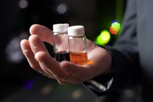
Anton Naumov holds bottles of carbon nanotubes, left, and quantum dots — two examples of nanomaterials used for biological imaging. Photo by Jeffrey McWhorter
Graphene was isolated in 2004 by physicists Andre Geim and Konstantin Novoselov of the University of Manchester in the United Kingdom. They pioneered a technique of using Scotch tape to isolate single-atom-thick layers from graphite and shared a Nobel Prize in 2010.
Naumov began working with graphene’s oxygen-laced version over a decade ago as a graduate student at Rice University in Houston, where he helped discover its acidity-dependent fluorescent properties with chemist R. Bruce Weisman.
Quantum dots could emerge as pioneering theranostics, or agents that can both locate a cancer and treat it, Weisman said.
Such an agent “would be injected into a patient, would find tumors and would, either directly or after activation, go after them,” Weisman said. “Multifunctionality is really one of the appeals of these nanoparticles, and [Naumov is] trying to exploit that.”
Naumov is now planning studies in mice to explore toxicity, magnetic targeting and tumor treatment. A decade might pass before quantum dots appear in a medical clinic, he estimated. But quantum dots’ versatility makes them an excellent bet.
“I believe that new nanomaterial-based platforms for cancer therapy should be multifunctional,” Naumov said. “This is the only way to effectively treat cancer because it is such a complex condition — it is such a variable and adaptable condition.”
Your comments are welcome
Comments
Related reading:
Features, Research + Discovery
Engineering a Possible Diabetes Treatment
Lauren Getz is working to coat pancreatic islet cells in hopes of improving treatment of Type 1 diabetes.
Alumni, Features
Making a Difference Through Medical Nonprofits
Four alumni physicians have a heart for healing and work to make the world a better place — one patient at a time.
Features
Ann Marie Herbst’s Matchmaking Mission
Now in remission from leukemia, the alumna aims to raise awareness for the bone marrow donor registry.