Photo by Rodger Mallison
Scientists See Medical Breakthroughs From Fluorescence
A new book shines a light on technology that can diagnose concussions faster.
In the mid-1600s, Nicolás Monardes, a Spanish physician and botanist, noticed a “bluish opalescence” emanating from a mixture of water and the pulp of a Mexican kidneywood tree.
In 1877, several kilograms of fluorescein, a compound using the same opalescence phenomenon, were poured into the Danube River to prove that it and the Rhine River were connected via underground waterways.
Today, the use of fluorescence has gone from underground streams to the human bloodstream.
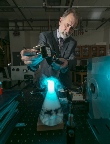
Karol Gryczynski, professor of physics in TCU’s College of Science and Engineering, studies fluorescence and its many uses. Photo by Rodger Mallison
Zygmunt “Karol” Gryczynski, the W.A. “Tex” Moncrief, Jr. Chair of Physics in the department of physics and astronomy at TCU and a professor in the department of microbiology, immunology and genetics at the University of North Texas Health Science Center, uses fluorescence to research innovative technologies, including biomedical advancements.
When Gryczynski started working with fluorescence, “it was kind of an exotic science,” he said. “Over time, it has become one of the major tools in biomedicine.
“I have been working in the biomedical field for a long time, and I naturally look for any applications of fluorescence technologies to biomedical diagnostics.”
The physicist has worked alongside Stanley Prusiner, Brian Kobilka and Stefan Hell, Nobel Prize winners who have all been involved in the field of fluorescence.
But one of Gryczynski’s closest scientific partnerships is with his older brother Ignacy Gryczynski, a professor in the department of microbiology, immunology and genetics at the UNT Health Science Center since 2005.
Working with his brother “helps a lot,” the younger Gryczynski said. “We don’t have to talk to each other a lot to understand what we want to do. Many things can be done faster.”
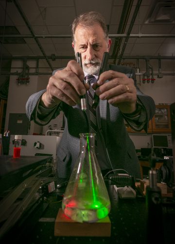
Ignacy Gryczynski’s tools can measure in nanoseconds the changes in the speed of the light traveling through test substances. Photo by Rodger Mallison
Ignacy Gryczynski, who is 10 years older, moved to the U.S. from Poland in the mid-1980s to further his research. He spent 20 years in Baltimore in the Center for Fluorescence Spectroscopy at the University of Maryland exploring a variety of applications for fluorescence.
Karol Gryczynski, who immigrated to the U.S. in 1989, has spent most of his career working with fluorescence, saying his brother “kind of dragged me into [the field].”
The brothers have co-published hundreds of scientific reports and book chapters, and they share more than a dozen patents.
The Gryczynski brothers’ complementary personalities give their partnership the tools to tackle most research situations. According to students, Ignacy Gryczynski said, Karol “is more like a mentor, and I am more like a friend.”
“They’re each kind of the same person but put through different personalities,” said Joe Kimball, a postdoctoral fellow who focuses on fluorescence and biomedical imaging. He works with the brothers on fluorescence-based projects.
“Ignacy is a lot more quiet; Karol is more loud,” Kimball said. “Ignacy speaks slower. Karol speaks faster. They’re both always in the lab and always working.”
New Book, New Experiments
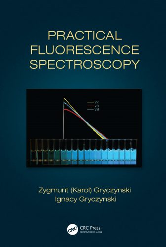
The Gryczynski brothers’ latest collaboration is an introductory book focused on fluorescence experiments. The publication is designed for new doctoral students as well as veteran researchers.
Courtesy of CRC Press | Taylor & Francis Group
Practical Fluorescence Spectroscopy (CRC Press | Taylor & Francis Group), published in late 2019, is a compilation of about 150 experiments, ranging from basic observations to advanced applications.
“Readers may be somewhat surprised that this book contains only a few original references,” the Gryczynskis write in the book’s introduction. “This is because all experiments were done by us and our students from the beginning to the end.”
With experiments that span several years, the brothers hope their book will contribute to the scientific renown of fluorescence spectroscopy. “This book was a 10-year idea,” Ignacy Gryczynski said, adding that the original concept was a short book on a few basic experiments.
Real-world examples give readers a more universal understanding of the material, such as how Rayleigh scattering, a form of light dispersal in which the light’s wavelength remains unchanged, is responsible for the blue color of the sky.
By the book’s last chapter, the professors provide more sophisticated research. One experiment lays out the procedure to follow the interaction between a fluorophore, a chemical compound that gives off fluorescence when subjected to light, and another molecule in solvent when they are brought together.
It is difficult to gather precise information from this interaction because, the book says, there are “many theories describing fluorophore-solvent interaction,” but there is “not [any] existing satisfactory theory of liquids.”
The experimental approach presents a possible workaround by applying research tactics outlined in earlier chapters. This combination of equipment and skills results in the ability to analyze and record the interaction between the fluorophores and biomolecules on a microscopic molecular scale.
While Ignacy Gryczynski wants to get started on the next book project, his brother isn’t quite ready. “I want to get [this book] over with, and then I have to rest for a few months, and then we can think about [the other book],” Karol Gryczynski said.
Leading to a New Device
In his TCU lab, Karol Gryczynski is working on several projects that involve fluorescence imaging and diagnostic methods. One in particular would make it faster and easier to diagnose concussions.
He wants to develop a portable tester that can track a person’s blood composition through fluorescence spectroscopy to determine if a concussion has occurred.
“I do not think there is a lot about portable fluorescence-based detection of concussive injuries or in fact any other portable detection of concussion,” he said.
Currently, concussions are detected through X-rays, MRIs or lumbar punctures (better known as spinal taps). These methods are expensive and invasive, and patients and samples must be tested off-site. Thus, only the most severe injuries are tested, but a concussion can occur in even minor incidents that are often ignored.
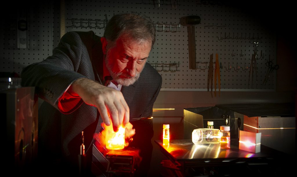
A current research project by Karol Gryczynski, the W.A. “Tex” Moncrief, Jr. Chair of Physics, might employ fluorescence to better diagnose concussions. Photo by Rodger Mallison
Gryczynski is working to develop a fluorescence compound that will identify the proteins released in brain cells after injury that diffuse into the bloodstream.
Past studies have shown that during a concussion, most of the proteins responsible for impaired cognitive function enter the bloodstream and tissue through leakage from the blood brain barrier.
With the right fluorescent marker, Gryczynski hopes to detect the presence and amount of the protein in the bloodstream. He expects that the portable tester will build a profile of a person’s blood composition over time to see how much of this protein is naturally present. That data would let medical personnel identify irregular spikes in the quantity of that protein, which in theory would suggest a concussion has occurred.
“If we can detect concussion markers in a drop of blood like in a glucose test,” he said, “then you can test the [patient] before and after the game with a prick of the finger.”
The portable tester would use fluorescence imaging, where a fluorescent marker is mixed into the blood sample and then subjected to a focused light excitation. The resulting image is, at its best, capable of distinguishing individual molecules of interest from the background.
“One very important aspect of fluorescence is that we gain very high sensitivity imaging due to the way molecules interact with different colors of light,” Gryczynski said. “The molecule absorbs the photon [light], holds the energy of the photon for a few nanoseconds and then releases the photon … of different wavelengths.”
Gryczynski is working to develop an effective device by increasing the sensitivity of the fluorescent compound and by perfecting the pulse imaging process used to illuminate the sample.
The proteins that indicate a concussive injury constitute just a few parts per billion of a person’s overall blood composition. Their scarcity makes it difficult to track any change in saturation.
To make more accurate measurements, Gryczynski is developing methods to increase the brightness of fluorescent markers that will respond with greater intensity when in contact with the proteins, making them more identifiable.
To accomplish this, the fluorophores are designed in such a way to treat the water-repellent and water-absorbent pockets of a specific protein as binding sites.
“The thing here that makes TCU different from other universities is that it’s not very big. … Organized collaboration with other departments is very easy. That’s what makes TCU great.”
Karol Gryczynski
Once bound, the fluorophore will emit an identifiably different fluorescent signal from its unbound kin, most often due to the compound changing from the polar (water-absorbent) environment of the solvent to the hydrophobic (water-resistant) state of the protein pocket.
Think of the fluorophore as a match. When in the wet part of the sample, the fluorophore ignites with less intensity or not at all because of the water saturating it. When in the dry pocket of a protein, however, the fluorophore can burn with greater power.
This grants the ability to detect not only the presence of a certain protein, but also its concentration in a given sample.
Another goal is to increase image brightness and resolution. Gryczynski plans to use a method similar to the brothers’ patented imaging process that involves multiple pulses of photons and emissions using special dyes developed with his collaborators at the University of Copenhagen.
In that process, a series of light pulses of a given wavelength excite the sample. Every pulse afterward increases the population of excited molecules used to detect the presence and concentration of components within the sample. This increases fluorescence spectroscopy’s potential for use in the medical field.
For concussion detection, the method helps to create a brighter picture that can better highlight the proteins of interest and lessen the background signal of the image.
“If you think about the human body,” Gryczynski said, “you have to realize everything we have is essentially a lot of garbage. Anything we do with fluorescence will detect this garbage or unwanted [background] signal.”
Although the proteins being tracked by the fluorescent compound will remain illuminated for a longer period of time, the difference is no more than a handful of nanoseconds. With only a brief window of time when just the target proteins will be illuminated, it is difficult to generate a clear picture of the sample.
To filter out the background signals and sharpen the image, the sample undergoes excitation with bursts of photons, similar to how the flash on a camera brightens and sharpens a photograph’s center of focus.
Calculating the most effective space between strobe pulses is vital to create the clearest possible image. If the pulse rate is too low, the wait for a clear image might allow the sample to change and make the image worthless. If the rate is too high, other parts of the sample aside from the markers of interest will remain illuminated and end up overexposing the background.
Through a measured barrage of light, the active fluorescent particles will become brighter while the less active background signal will remain dim, improving overall image brightness and resolution and creating a picture that can identify concussion indicators in the bloodstream.
An Environment of Collaboration
From the University of Strathclyde in Scotland to Shimane University in Japan, Gryczynski travels often as a visiting professor. But TCU is where he feels empowered to pursue his goals.
“The thing here that makes TCU different from other universities is that it’s not very big,” he said. “What that means is that everyone on campus knows each other, so organized collaboration with other departments is very easy. That’s what makes TCU great.”
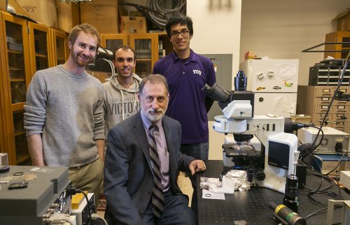
Karol Gryczynski, seated, works with research associate Joe Kimball and graduate students Luca Ceresa and Jose Chavez, from left, as he studies fluorescence and its many uses in his laboratory in the Sid Richardson Building at TCU. Photo by Rodger Mallison
Gryczynski encourages his students to take advantage of TCU’s capacity to create connections with experts from different professions, from physicians to business leaders.
“We have met so many people,” Kimball said. “Easily 200 people from across the world have visited this lab while I’ve been working here. We’re always doing something new from someone’s new idea.”
Gryczynski has integrated his research into the classroom, but he has bigger ideas. He wants to spread fluorescence and physics to every corner of the university. “I [am] at that point in my career where I [want] to make physics accessible to everybody,” he said.
“I think it is an important science. It gives a lot of potential in application.”
Robert Pendry ’15, who was in Gryczynski’s biomedical imaging class in Fall 2013, recounts how the course engaged students from other majors. “He’s making a really big effort in making everything translational,” Pendry said. “His classes aim to do that by presenting the science, but then also saying, ‘If you’re a doctor or a radiologist, and you had to use this imaging technology, why would it be preferable to other imaging technologies?’ ”
Pendry earned a double degree, in physics and astronomy and in neuroscience, at TCU. Today he is a doctoral student in the department of neuroscience at UT Southwestern Medical Center in Dallas. He is also a guest lecturer for the same biomedical imaging class he took from Gryczynski.
The professor would like to develop a course with the Neeley School of Business on entrepreneurship that would cover a technology’s development to commercialization and bring together students from different areas of study. He said that while he and many other scientists know how to develop and patent new technologies, they are less familiar with marketing products derived from their research.
“I want to put these patents to use,” Gryczynski said. “If we develop a collaboration with the business school, it could be attractive to a lot of students looking to develop a business.”
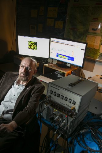
As part of his research at the UNT Health Science Center, Ignacy Gryczynski uses a microscope equipped with lasers and sophisticated sensors. Photo by Rodger Mallison
Michael Sherrod, William M. Dickey Entrepreneur in Residence at the TCU Neeley Institute for Entrepreneurship and Innovation, encourages this collaboration. “Where the Neeley School would be really useful is helping to transfer patents into a business opportunity … and [we] are eager for this interaction with other parts of the campus.”
With this collaboration and more in mind, Gryczynski hopes to show others the value of physics and fluorescence and how these sciences can benefit many disciplines.
With their new book, the brothers aim to further this cause by creating an accessible, comprehensible and in-depth analysis of experiments in fluorescence spectroscopy. “If we make physics interesting to others outside the major,” Gryczynski said, “then I consider it a success.”
Karol Gryczynski’s research is made possible by the W.A. “Tex” Moncrief, Jr. Endowed Chair of Physics.
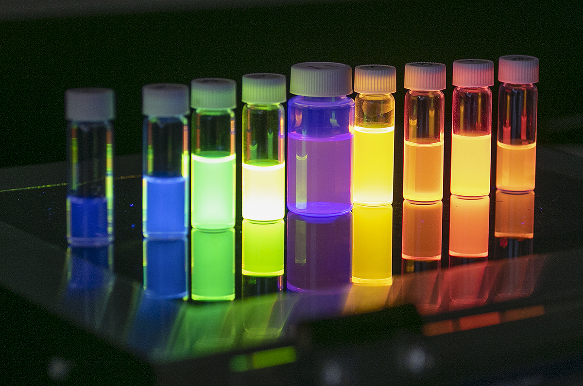
What Is Fluorescence?
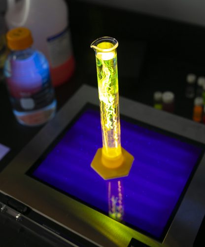
Photo by Rodger Mallison
The term fluorescence was coined by Sir George Stokes in the 1850s after he observed the phenomenon in a chunk of fluorspar. It is the process whereby a substance absorbs light from an external source and emits it at a longer wavelength. For this to occur, the substance must either be or contain a fluorophore.
Fluorophores are molecules capable of absorbing light energy within a specific wavelength range. That energy excites the electrons within the molecule and makes them unstable. To return the molecule to a stable state, the electrons release the light energy at a longer wavelength.
Think of the fluorophore as a punching bag. The punching bag absorbs the energy from the punches and disperses that energy out across its entire body at a lower velocity, converting a knockout right hook into little more than a gentle shove.
— Nicholas Ferrandino
Sources: Shannon Luminous Materials (blacklite.com) and Science Squared (bitesizebio.com)
How Does Fluorescence Microscopy Work?
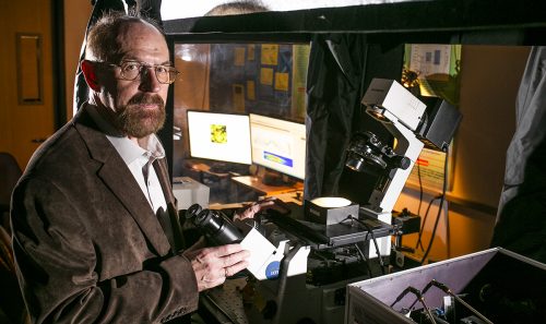
Karol Gryczynski investigates fluorescence as a tool in biomedicine. Photo by Rodger Mallison
The fluorophore’s ability to lengthen the wavelength of an external light source makes it possible to see light frequencies such as X-rays that are otherwise invisible to the naked eye.
Fluorescence spectroscopy and other forms of fluorescence imaging take full advantage of this by controlling both the fluorophore and the light source to study samples in greater detail.
To start, fluorescent dyes or proteins are mixed into a sample, where they will bond to a specific molecule or compound and become active.
The sample is then put under a specialized microscope that shoots it with an adjustable beam of light, exciting the activated fluorophores and highlighting the substance that the dye bonded to. This makes fluorescence imaging devices adept at tracking and measuring specific compounds along with their patterns and characteristics as they become brighter than the inactive areas of the sample.
— Nicholas Ferrandino
Source: ThermoFisher Scientific
Your comments are welcome
Comments
Related reading:
Research + Discovery
A Silver Bullet for Cancer Treatment?
Nanoparticles may be able to tame an effective but damaging chemotherapy medication.
Campus News: Alma Matters, Research + Discovery
Altering Antibiotics
Student in TCU high school program works on medicines of tomorrow.
Research + Discovery
Eclipsing Research
TCU astronomer Kat Barger shines a light on the Milky Way’s origins.